

Sometimes your body whispers before it ever shouts. Maybe it’s a heaviness after meals, unusual bloating, unexplained fatigue, or a feeling you can’t quite put your finger on. With liver-related issues, symptoms often stay vague until the condition becomes more serious.
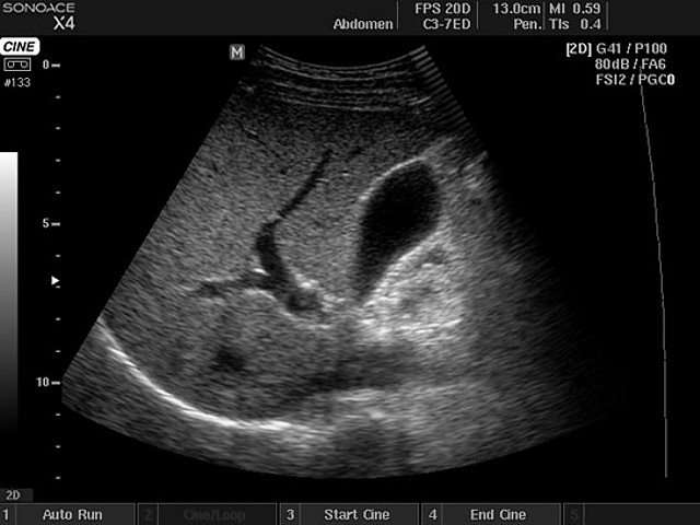
A liver ultrasound (also known as a liver scan) is one of the most reliable, gentle, and non-invasive ways to understand what’s happening inside long before problems escalate. During the procedure, real-time images of your liver and surrounding structures are displayed on a computer screen for immediate analysis. At MyConciergeMD in Los Angeles, we offer this test to give you clarity, comfort, and answers rooted in real data, not guesswork.
What the liver does – and why it’s essential
Your liver is one of the hardest-working organs in your body. It:
- Filters toxins and chemicals
- Processes medications
- Helps digest fats
- Stores vitamins and essential nutrients
- Regulates metabolism and blood sugar
- Produces proteins that help your blood clot
The liver is a key part of the biliary system, which includes the gallbladder and bile ducts, and is responsible for producing and transporting bile to aid in digestion.
When the liver is strained or unhealthy, everything, from digestion to energy levels, can be affected. Because many liver diseases develop silently, early imaging with liver ultrasound is key to staying ahead of potential issues.
Why doctors recommend a liver ultrasound
Doctors may recommend a liver ultrasound if you experience or have:
- Abdominal pain or discomfort in the right upper quadrant
- Fatty liver disease (NAFLD) or alcohol related liver disease
- Elevated liver enzymes (ALT, AST, ALP)
- Hepatitis B or C
- Chronic hepatitis (such as hepatitis B or C)
- Jaundice or yellowing of the skin
- Unexplained bloating or fluid retention
- Liver cysts, nodules, or tumors
- History of alcohol use
- Gallbladder disease or biliary obstruction
- Unexplained weight loss or appetite changes
An upper right quadrant ultrasound focuses on the liver and nearby structures, and may also visualize other organs such as the gallbladder, pancreas, and right kidney.
It is also commonly performed as part of routine abdominal imaging or annual wellness assessments.
Preparation for the liver ultrasound
To get the most accurate images:
- Fast for 6–8 hours before your appointment. Fasting helps reduce gas and ensures the liver and gallbladder can be seen clearly, as intestinal gas can block the ultrasound beam and make it harder to obtain clear images.
- Avoid heavy meals the night before
- Take medications as usual unless told otherwise
- Drink water only if needed
- Avoid carbonated beverages and smoking before the scan (these introduce air in the abdomen)
Procedure of liver ultrasound at My Concierge MD
A liver ultrasound typically takes 15-25 minutes and is completely painless. Here’s what you can expect:
- You’ll lie on your back or slightly turn to the left.
- A warm gel is placed over your right upper abdomen by our ultrasound technician.
- The ultrasound technician performs an ultrasound of the liver, focusing on the upper right quadrant (quadrant ultrasound), and moves the ultrasound probe along your ribs and upper abdomen.
- Images of the liver, gallbladder, and nearby structures are captured.
After the scan, a detailed report (ultrasound report) is prepared by the radiologist, summarizing the findings for your doctor.
What Liver Ultrasound Scan Confirms?
- Liver size and texture
- Fatty infiltration (fatty liver)
- Echogenic liver (increased brightness indicating possible fatty liver, hepatitis, or cirrhosis)
- Cysts, tumors, or nodules
- Solid masses (distinguishing tumors from cysts or fluid collections)
- Inflammation or signs of chronic liver disease
- Dilated bile ducts (suggesting possible blockages or obstructions)
- Blockages in the bile ducts
- Gallstones or gallbladder disease
- Comparison to a normal liver appearance
Liver Cancer Detection with Ultrasound
Detecting liver cancer early can make all the difference in treatment and outcomes. Liver ultrasound uses high-frequency sound waves to create detailed images of the liver, making it an essential tool for identifying liver lesions, tumors, and other abnormalities. With the addition of Doppler ultrasound, doctors can assess blood flow within the liver and its major vessels, which helps distinguish between benign and malignant lesions. This vascular liver ultrasound approach is especially valuable for spotting signs of hepatocellular carcinoma (HCC), the most common type of primary liver cancer.
For individuals with chronic liver disease, cirrhosis, or a history of hepatitis B or C, regular liver ultrasound screening is recommended. This imaging test is non-invasive, does not involve radiation exposure, and can detect subtle changes in liver tissue before symptoms appear. By monitoring blood flow and liver structure, ultrasound can help catch liver cancer at an early, more treatable stage, providing peace of mind and a clear path forward for those at risk.
Fatty Liver Diagnosis and Assessment
Fatty liver disease, or hepatic steatosis, is increasingly common and often silent in its early stages. Liver ultrasound is a frontline imaging test for detecting and assessing fatty liver. During the scan, areas of increased echogenicity (brightness) signal the presence of fatty tissue within the liver. The degree of brightness can help estimate the severity of fatty liver disease, and advanced techniques like elastography ultrasound can measure liver stiffness—a key indicator of fibrosis or scarring.
Elastography ultrasound is particularly useful for monitoring the progression of fatty liver disease and evaluating the risk of developing more serious liver conditions. While a liver biopsy may still be needed for a definite diagnosis in some cases, ultrasound offers a safe, non-invasive, and cost-effective way to screen for and track fatty liver disease over time.

